Embark on an enlightening journey through Brain and Cranial Nerves Lab 26, where we unravel the intricate tapestry of the human brain and its vital connections to the body. Join us as we delve into the depths of neuroanatomy, exploring the brain’s remarkable structures and the crucial role of cranial nerves in orchestrating our sensory, motor, and cognitive functions.
Prepare to be captivated as we navigate the complexities of the brain, identifying its major regions and deciphering their diverse functions. We will trace the pathways of the 12 pairs of cranial nerves, unraveling their intricate distributions and understanding their indispensable contributions to our ability to perceive, interact, and thrive.
Brain and Cranial Nerves Lab 26 Introduction
Lab 26: Brain and Cranial Nerves is designed to provide you with a comprehensive understanding of the anatomy and function of the brain and cranial nerves. Through hands-on dissections and interactive exercises, you will gain valuable insights into the complex structures and pathways involved in brain function and sensory perception.
The brain is the central organ of the nervous system, responsible for coordinating and controlling various bodily functions, including movement, sensation, cognition, and emotion. Cranial nerves are 12 pairs of nerves that originate from the brain and innervate structures in the head and neck, such as muscles, glands, and sensory organs.
Objectives
- Identify the major structures of the brain and describe their functions.
- Trace the pathways of cranial nerves and understand their sensory and motor functions.
- Correlate the anatomy of the brain and cranial nerves with clinical presentations.
- Apply your knowledge to solve clinical case studies involving brain and cranial nerve disorders.
Materials and Methods
The Brain and Cranial Nerves Lab 26 requires various materials to facilitate the exploration of the brain and its associated cranial nerves. These materials include:
- Preserved sheep brain
- Dissecting tools (scalpel, scissors, forceps)
- Magnifying glass or dissecting microscope
- Charts or diagrams of the brain and cranial nerves
- Preserved cranial nerves (optional)
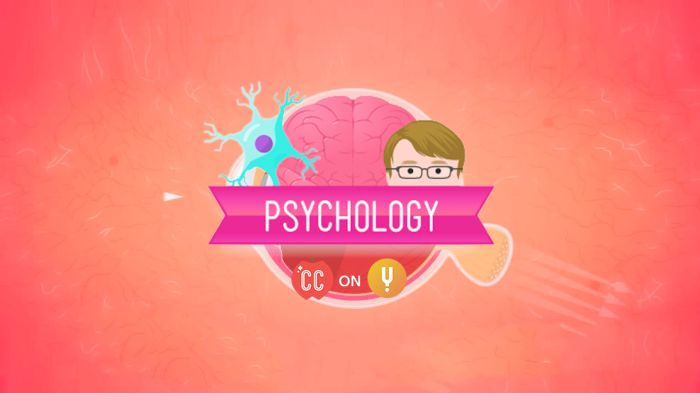
The lab procedures involve a series of steps to examine the brain and cranial nerves. Firstly, the preserved sheep brain is placed in a dissecting tray and carefully observed. Using dissecting tools, students make incisions and dissections to reveal the internal structures of the brain, including the cerebrum, cerebellum, and brainstem.
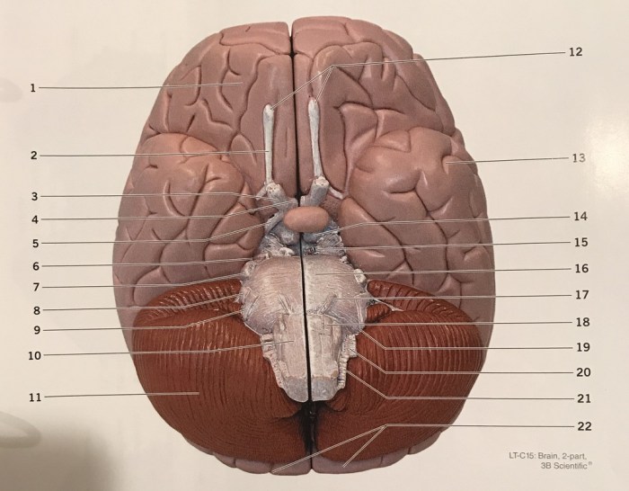
External Brain Anatomy
The external anatomy of the brain is examined, including the major lobes, sulci, and gyri. Students identify and locate key structures such as the frontal lobe, parietal lobe, occipital lobe, temporal lobe, and cerebellum.
Internal Brain Anatomy
Internal dissections are performed to reveal the internal structures of the brain. Students observe the ventricles, thalamus, hypothalamus, and other deep brain structures. The connections between different brain regions are also examined.
Cranial Nerves
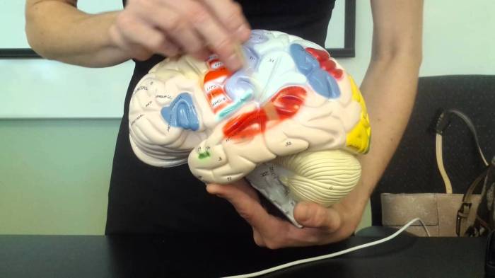
The cranial nerves are identified and traced from their origins in the brain to their distribution in the head and neck. Students observe the location, function, and distribution of each cranial nerve, gaining an understanding of their role in sensory and motor functions.
Brain Anatomy: Brain And Cranial Nerves Lab 26
The brain is the control center of the nervous system and is responsible for a wide range of functions, including thought, emotion, movement, and memory. It is divided into three main regions: the forebrain, midbrain, and hindbrain.
The forebrain is the largest and most complex region of the brain. It is responsible for higher-level functions such as cognition, language, and memory. The forebrain is divided into two hemispheres, the left and right hemispheres, which are connected by the corpus callosum.
The midbrain is a small region of the brain that connects the forebrain to the hindbrain. It is responsible for controlling eye movements and coordination.
The hindbrain is the most posterior region of the brain. It is responsible for controlling basic life functions such as breathing, heart rate, and blood pressure. The hindbrain is divided into two regions, the medulla oblongata and the pons.
Major Structures of the Brain
- Cerebrum:The cerebrum is the largest part of the brain and is responsible for higher-level functions such as cognition, language, and memory.
- Cerebellum:The cerebellum is located at the back of the brain and is responsible for coordination and balance.
- Brainstem:The brainstem is located at the base of the brain and is responsible for controlling basic life functions such as breathing, heart rate, and blood pressure.
- Thalamus:The thalamus is located at the base of the cerebrum and is responsible for relaying sensory information to the cerebrum.
- Hypothalamus:The hypothalamus is located at the base of the cerebrum and is responsible for regulating body temperature, hunger, thirst, and sleep.
Cranial Nerves
The cranial nerves are 12 pairs of nerves that originate from the brain and extend to various parts of the head, neck, and trunk. They are responsible for a wide range of functions, including sensory perception, motor control, and autonomic regulation.
The 12 pairs of cranial nerves are:
- Olfactory nerve: Responsible for the sense of smell.
- Optic nerve: Responsible for vision.
- Oculomotor nerve: Controls the movement of the eye muscles, except for the superior oblique muscle.
- Trochlear nerve: Controls the movement of the superior oblique eye muscle.
- Abducens nerve: Controls the movement of the lateral rectus eye muscle.
- Facial nerve: Controls the muscles of facial expression.
- Vestibulocochlear nerve: Responsible for hearing and balance.
- Glossopharyngeal nerve: Responsible for taste sensation in the posterior third of the tongue and motor control of the pharynx.
- Vagus nerve: Responsible for sensation and motor control of the pharynx, larynx, and thoracic and abdominal viscera.
- Accessory nerve: Responsible for motor control of the sternocleidomastoid and trapezius muscles.
- Hypoglossal nerve: Responsible for motor control of the tongue.
li> Trigeminal nerve: Responsible for sensation in the face and motor control of the muscles of mastication.
The cranial nerves are essential for a wide range of functions, and damage to any of these nerves can lead to significant neurological deficits.
Case Studies and Clinical Applications
Case studies and clinical applications provide valuable insights into the clinical relevance of the brain and cranial nerves. Understanding the anatomy and function of the cranial nerves enables healthcare professionals to effectively diagnose and treat neurological disorders.
Lab 26 on the brain and cranial nerves is quite the handful, but hey, who said learning about the nervous system couldn’t be a little puzzling? If you’re stuck on a crossword clue asking for a 7-letter word for “behind,” check out this to get back on track.
Then, you can dive right back into the fascinating world of neuroanatomy in lab 26.
A cranial nerve examination is a crucial component of a neurological assessment. By examining the function of each cranial nerve, clinicians can assess the integrity of the brain, brainstem, and peripheral nervous system. Abnormalities in cranial nerve function can indicate various neurological conditions, including:
Cranial Nerve Palsies
- Unilateral or bilateral weakness or paralysis of specific muscles innervated by a particular cranial nerve.
- May result from lesions in the brain, brainstem, or peripheral nerves.
- Can manifest as difficulty with eye movements, facial expressions, swallowing, or speech.
Increased Intracranial Pressure
- Papilledema (swelling of the optic nerve head) due to elevated intracranial pressure.
- May indicate conditions such as brain tumors, meningitis, or hydrocephalus.
- Cranial nerve VI (abducens) palsy is a common finding associated with increased intracranial pressure.
Neurodegenerative Disorders
- Cranial nerve involvement is a common feature in neurodegenerative disorders like Parkinson’s disease and Alzheimer’s disease.
- Progressive damage to cranial nerve nuclei or pathways can lead to impaired eye movements, speech, and swallowing.
- Early detection and management of cranial nerve dysfunction can improve patient outcomes and quality of life.
Data Analysis and Interpretation

Analyzing and interpreting the data collected during the lab is crucial to understanding brain function. The data should be evaluated to identify patterns, trends, and relationships between different brain structures and their associated functions.
The significance of the findings lies in their implications for understanding how the brain processes information, controls movement, and regulates bodily functions. By analyzing the data, researchers can gain insights into the neural mechanisms underlying various cognitive, motor, and sensory processes.
Statistical Analysis
Statistical analysis is employed to determine the significance of the observed data. Statistical tests, such as t-tests or ANOVAs, can be used to compare the performance of different brain structures or the effects of different experimental conditions.
Correlation and Regression Analysis, Brain and cranial nerves lab 26
Correlation and regression analysis can reveal relationships between different brain structures or functions. Correlation analysis measures the strength and direction of the relationship between two variables, while regression analysis can predict the value of one variable based on the values of other variables.
Neuroimaging Techniques
Neuroimaging techniques, such as fMRI and EEG, can provide valuable data for analyzing brain function. These techniques allow researchers to visualize brain activity and measure changes in brain structure and function in response to various stimuli or tasks.
Clinical Applications
The findings from brain and cranial nerves research have significant clinical applications. By understanding the neural mechanisms underlying various neurological disorders, researchers can develop more effective treatments and interventions.
Conclusion
The brain and cranial nerves lab provided a comprehensive overview of the anatomy and function of the brain and cranial nerves. Through hands-on dissection and observation, we gained a deeper understanding of the complex structures and pathways involved in sensory and motor function.
The key findings of the lab include:
- Identification of the major structures of the brain, including the cerebrum, cerebellum, brainstem, and ventricles.
- Tracing the pathways of the cranial nerves and understanding their sensory and motor functions.
- Appreciation for the intricate connections between the brain and the rest of the body.
While the lab provided valuable insights, it also highlighted the limitations of our current understanding of the brain and cranial nerves.
Limitations and Future Research
One limitation of the lab was the use of preserved specimens. While this allowed for detailed dissection and observation, it did not provide a complete picture of the functioning brain. Future research could focus on developing non-invasive imaging techniques that allow for the study of the brain in vivo.
Another limitation was the limited time available for dissection. As a result, we were not able to explore all aspects of the brain and cranial nerves in depth. Future research could involve more extensive dissections or the use of virtual reality simulations to provide a more comprehensive understanding.
Despite these limitations, the brain and cranial nerves lab was a valuable learning experience. It provided a foundation for further study of the nervous system and highlighted the importance of continued research in this field.
Detailed FAQs
What is the purpose of Brain and Cranial Nerves Lab 26?
Brain and Cranial Nerves Lab 26 provides an in-depth exploration of the brain’s anatomy and the functions of the 12 pairs of cranial nerves. It offers hands-on experience in identifying and understanding these vital structures and their roles in sensory, motor, and cognitive functions.
What materials are required for the lab?
The lab requires various materials, including preserved brain specimens, dissection tools, anatomical charts, and reference materials. Specific requirements may vary depending on the institution and the scope of the lab.
How are the data collected during the lab analyzed and interpreted?
Data collected during the lab, such as observations and measurements, are analyzed to identify patterns and draw conclusions about brain anatomy and cranial nerve functions. Statistical methods may be employed to determine the significance of findings and assess the validity of hypotheses.